The NICHD 17th Annual Meeting of Postdoctoral, Clinical, and Visiting Fellows, and Graduate Students and Postbacs took place virtually on September 29, 2022. Dr. Henry Levin, Senior Investigator in the Section on Eukaryotic Transposable Elements, opened the day with a big thank you to the NICHD Office of Education and retreat steering committee. He then commented on the pandemic and its impact on the research and mental wellness of NIH fellows before offering a celebration of recent NICHD trainee scientific accomplishments (see our 2022 Year in Review!).
Following Dr. Levin’s opening remarks, fellows were treated to a day of learning and career exploration. The day included two insightful keynotes, a review of innovative culture at NICHD, multiple career focus panels, and engaging presentations from 27 trainees (including 16 three-minute poster talks).
Please enjoy the following recap of the morning keynote, the NICHD Innovative Culture Initiative introduction, and the featured fellow presentations from the 2022 Virtual Fellows Retreat—all written by NICHD trainees.
The 2022 Retreat Steering Committee
Chair: Anna Santamaria, PhD (postdoc, Rouault Lab)
- Hyo Won Ahn, PhD (postdoc, Levin Lab)
- Avik Dutta, PhD (postdoc, Love Lab)
- Henry Lessen, PhD (former postdoc, Sodt Lab)
- Amrita Mandal, PhD (postdoc, Balla Lab)
- Thien Nguyen, PhD (postdoc, Gandjbakhche Lab)
- Jeremie Oliver (graduate student, D’Souza Lab)
- Tusharkumar Patel, PhD (postdoc, Yanovski Lab)
- Christina Porras, PhD (postdoc, Rouault Lab)
- Megha Rajendran, PhD(postdoc, Bezrukov Lab)
- Bo-Mi Song, PhD(postdoc, Stopfer Lab)
- Sanjana Sundararajan, PhD (postdoc, Dasso Lab)
- Abhinav Sur, PhD(postdoc, Farrell Lab)
Decoding Signals, Developing Therapies
Lysosomes are more than degradative compartments, said Rosa Puertollano, PhD, Senior Investigator, Laboratory of Protein Trafficking and Organelle Biology, National Heart, Lung, and Blood Institute, during her keynote presentation at the fellow’s retreat. She emphasized throughout her talk that lysosomes have an important role in cellular stress response, signaling, and disease.
Dr. Puertollano detailed how extracellular signals pass through lysosomes, combine with cytosolic signals, and form signaling cascades that can regulate transcription inside the nucleus. For example, nutrient deprivation inhibits the lysosome localized mammalian target of rapamycin complex (mTORC1) and activates the transcription factor TFEB, upregulating multiple lysosomal and autophagy-related genes. In fact, the latest research from Dr. Puertollano’s lab places TFEB and TFE3 (another transcription factor) as important cogs in the cellular response to oxidative and DNA damage, as well as defense against pathogen infection.
Dr. Puertollano also presented research into Pompe disease, a lysosomal storage disorder caused by defects in the lysosomal enzyme Acid Alpha Glucosidase (GAA). This lysosomal disfunction ultimately leads to a buildup of aberrant mitochondria and autophagic vesicles, thereby starting a signaling cascade of cell death. Her research is tackling the disease by increasing the efficacy of enzyme replacement therapy while at the same time developing novel gene therapy methods.
Summarizing her career, Dr. Puertollano emphasized that planning early for the postdoctoral to principal investigator transition is critical, and she encouraged fellows to identify nascent fields where many fundamental discoveries can be made. Her keynote address showcased how fundamental research in cell and molecular biology can provide insights that lead to better therapies and cures for diseases. It was an inspiring story that set the tone for the rest of the retreat.
Cultivating an Innovative Culture at NICHD
The NICHD Innovative Culture Initiative (3.1.2) was conceived as a result of the Management and Accountability focus area of the NICHD Strategic Plan 2020. The core team proposed several definitions for innovation based on benchmarking and best practices. After polling senior leaders at NICHD, the Institute now defines innovation as “translating NICHD’s evolving needs and opportunities into new or improved services, processes, systems, or social interactions that promote an enhanced workforce, infrastructure, efficiency, and a culture that encourages continuous improvement through creativity and idea exploration.”
Benchmarking results and interviews from peer organizations suggest that focusing on four main elements—people, behavior/values, governance, and sustainability—may support successful development of an innovative culture. The two main aims of the initiative are (1) to reinforce a culture where staff feels empowered to propose solutions and communicate new ideas that will encourage continuous improvement and stimulate change, and (2) to develop processes and strategies for promoting innovative best practices that can be incorporated into the NICHD working environment.
In October of this year, the 2022 NICHD Innovative Culture Survey was distributed to all NICHD full-time staff and fellows to evaluate the extent to which the Institute’s current culture supports innovation. Building on the survey, focus groups will further clarify insights and feedback. Please reach out to NICHDInnovativeCulture@mail.nih.gov if you would like to volunteer to be part of a focus group! Participation is very welcomed!
Featured Fellow Talks
Mild Traumatic Brain Injury Induces Multiple Response Pathways in Cortical Neurons
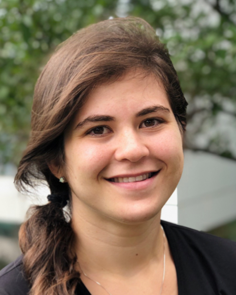
Mor Alkaslasi
Mild Traumatic Brain Injury has widespread effects on the brain, from blood vessel damage to the induction of glial responses, but it is unclear how these injuries affect the health of individual neurons. Mor Alkaslasi, a graduate student in the Le Pichon laboratory (Unit on the Development of Neurodegeneration), studies how neurons respond to this type of injury at the cellular level.
Using a mouse model with fluorescently tagged neurons that activate Activated Transcription Factor 3 (Atf3), a transcription factor expressed in injured neurons, Ms. Alkaslasi observed the morphology of sensory and motor cortex neurons following mild traumatic brain injury. While most Atf3-expressing neurons were undergoing apoptosis, the change in the number of Atf3-expressing neurons following mild traumatic brain injury differed across cortical layers and neuron type. Ms. Alkaslasi also conducted single-nucleus RNA sequencing and found differences in genetic expression that may be predictive of whether the neurons would die. Future work aims to elucidate the different pathways that are employed by neuron types in response to mild traumatic brain injury.
Characterization and Categorization of Transcriptional Trajectories During Zebrafish Development
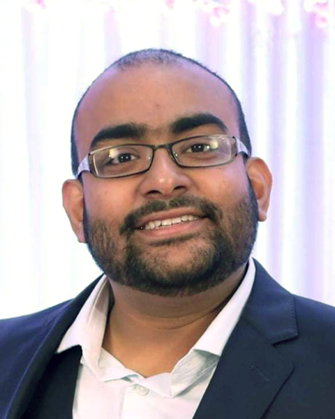
Abhinav Sur, PhD
With the vast number of cell types that generate during animal development, biological systems must coordinate this differentiation through programmed genetic expression. Abhinav Sur, PhD, a postdoctoral fellow in the Farrell laboratory (Unit on Cell Specification and Differentiation), seeks to characterize such gene expression programs through his work using single-cell transcriptomics and bioinformatic analyses.
Dr. Sur and his colleagues have created a high-resolution single-cell gene expression atlas of zebrafish development encompassing the first five days of embryogenesis. Using this atlas, they explored whether gene expression programs are shared across distinct tissues and have shed light on poorly understood cell types. Specifically, the team found a poorly understood cell type in the zebrafish intestine called best4+/otop2+ cells that were only recently discovered (in 2019) in the human intestine. These cells share several genes expressed in human counterparts. Dr. Sur has characterized the sequence of transcriptional changes underlying the development of many cell types including best4+/otop2+ cells. The team is currently developing an online resource, called Daniocell, to openly share this atlas with other researchers across the world.
A Medley of Metals and Proteins in Iron Sensing
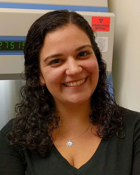
Anna SantaMaria, PhD
The production of red blood cells relies on a dance between regulatory proteins and iron. While iron is an element essential for many functions in the body including red blood cell production and oxygen transport, a high level of free iron causes toxicity and cell death. Anna SantaMaria, PhD, postdoctoral fellow in the Rouault lab (Section on Human Iron Metabolism), wants to understand how intracellular iron levels are sensed and controlled.
In cells, most iron atoms are stored in a large iron-storing protein called ferritin. When intracellular iron levels are high, a protein called Nuclear Receptor Coactivator 4 (NCOA4) prevents ferritin from being engulfed by autophagosomes, the cellular degradation system, and iron stays safely locked up. However, under iron starvation, NCOA4 promotes the degradation of ferritin-iron complexes allowing for the release of iron from storage. Dr. SantaMaria’s preliminary research suggests that NCOA4 may be binding iron-sulfur cluster(s) when iron levels are sufficient, thus allowing it to sense and react to iron levels. In the future, she plans to use spectroscopic techniques to detect the signatures of iron-sulfur clusters bound to NCOA4. Additionally, she aims to determine biological function of the putative iron-sulfur cluster by mutagenizing the iron-sulfur cluster binding residues in NCOA4 and observing the effect on iron metabolism and red blood cell production.
How Radiopharmaceuticals are Used to Detect Pheochromocytoma
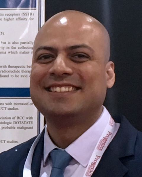
Abhishek Jha, MD
Abhishek Jha, MD, research fellow in the Pacak laboratory (Section on Medical Neuroendocrinology), studies the diagnostic performances of various radiopharmaceuticals in the detection of pheochromocytoma and paraganglioma. Pheochromocytomas (PHEOs) are rare neuroendocrine tumors originating from chromaffin cells of the adrenal gland, and paragangliomas are those that arise extra-adrenally. Normally found in adrenals or in groups of nerve cells called ganglia, chromaffin cells are responsible for producing neurotransmitters, such as adrenaline (epinephrine) and noradrenaline (norepinephrine), and releasing them into the blood stream. However, an overabundance of neurotransmitters from PHEOs can prove catastrophic for patients.
To determine the most sensitive diagnostic functional imaging modality in patients with PHEOs caused due to mutation in rearranged during transfection (RET), Dr. Jha compared positron emission tomography/computed tomography (PET/CT) scans using several radiopharmaceuticals (18F-FDOPA, 68Ga-DOTATATE, 18F-FDG, 18F-FDA) in 19 patients. Dr. Jha found that 18F-FDOPA had the highest detection rate, but due to the small number of patients, he emphasized that a larger multicentric study is warranted.
How Zebrafish Repair Blood Vessels
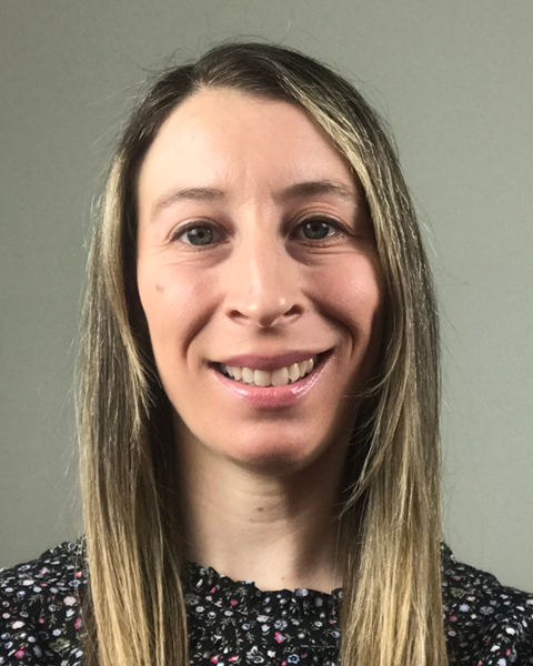
Leah Greenspan, PhD
Leah Greenspan, PhD, postdoctoral fellow in the Weinstein laboratory (Section on Vertebrate Organogenesis), investigates the cellular behaviors and molecular mechanisms driving blood and lymphatic vessel repair after injury. Her latest work suggests that signaling mechanisms differ between initial vessel patterning versus vessel healing.
Using transgenic adult fish, Dr. Greenspan delivered skin-deep cuts and visualized blood and lymphatic vessel regrowth through high-resolution confocal microscopy. While the injury completely healed after ten days, vessel patterning didn’t return to its preinjury state. To examine how endothelial cells move during injury, Dr. Greenspan induced cell death in a small subset of vessels in larval zebrafish. While most blood vessels reconnected normally due to endothelial cell migration, some vessels adopted a new identity, demonstrating the plasticity of vessels after injury. As a next step, Dr. Greenspan plans to analyze gene expression changes during damage and recovery.
On Our Way to a New Preclinical Model for Juvenile ALS?
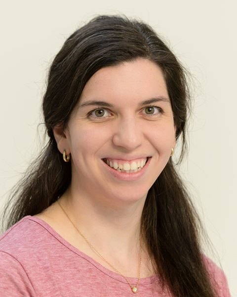
Zoe Piccus
Amyotrophic lateral sclerosis (ALS) is a fatal neurodegenerative disease, typically appearing around 40 to 50 years of age. Juvenile ALS onset, however, is before the age of 25. Juvenile ALS has been associated with mutations in an enzyme that initiates and regulates biosynthesis of sphingolipids, important membrane lipids with roles in development and cell function. These mutations are predicted to increase canonical sphingolipid levels. Zoe Piccus, a graduate student in the Le Pichon laboratory (Unit on the Development of Neurodegeneration), studies the connection between sphingolipid levels and disease pathology in a preclinical animal model of juvenile ALS.
Ms. Piccus generated a mouse model containing the juvenile-associated ALS mutation and assessed the mice for elevated sphingolipid levels and ALS-like neurodegeneration. Piccus found that circulating sphingolipids in serum harvested from mutant mice exhibited increased levels of canonical sphingolipids. The mutant mice also demonstrated a neurodegenerative phenotype characterized by age dependent increases in serum neural filaments, changes in nerve ultrastructure, signs of degenerating axons, decreases in nerve to muscle connectivity, and mislocalization of certain proteins—supporting its use as an animal model of juvenile ALS.
A Novel Player in Arterial Development
Miranda Marvel, PhD
Epigenetic modifications trigger large-scale, programmatic changes during development. Postdoctoral fellow Miranda Marvel, PhD, in the Weinstein laboratory (Section on Vertebrate Organogenesis) recently identified kdm4ab as a novel epigenetic regulator of arterial development in zebrafish.
Zebrafish development serves as a foundation for understanding human development, thus understanding epigenetic regulators of the circulatory system has direct implications on human health. Dr. Marvel used EpiTag transgenic zebrafish, which differentially express fluorescent proteins in response to changes in DNA or histone methylation, to link abnormal epigenetic patterns with defects in arterial vessel development. Looking for fish with abnormal arterial vessels, she zeroed in on a gene called kdm4ab. The kdm4ab mutant zebrafish exhibited downregulated arterial gene markers and upregulated venous marker genes, according to RNA-seq experiments. Dr. Marvel plans to further examine the association between downregulated arterial marker genes and altered histone methylation patterns to better assess kdm4ab’s epigenetic role.
The Beauty of (A)symmetry

Tyler Bruno
Structural asymmetries are essential across all stages of development. For example, asymmetries help orient cells along the plane of a tissue. Tyler Bruno, a postbaccalaureate fellow working in the Sackett laboratory (Cytoskeletal Dynamics Group), previously studied (with Dr. Becky Burdine at Princeton University) the role of a protein called Kurly in establishing these asymmetries during embryonic development. The role of Kurly in establishing developmental asymmetries in ciliated tissues has been extensively detailed, but Mr. Bruno wants to uncover Kurly’s role in development in nonciliated tissue contexts.
Mr. Bruno first explored the impacts of Kurly mutations on early zebrafish development. At early stages, Kurly mutants divided into disorganized clumps of cells—in contrast with the well-structured wild-type—but the mutant morphology normalized by the 256-cell stage. Bruno also characterized the downstream impacts of Kurly’s role in cellular asymmetry by measuring cellular shape and structure at both early and later developmental time stages. In all, Mr. Bruno hypothesized that Kurly serves as a scaffolding protein, recruiting other proteins that help initialize polarized cellular processes.
Just Keep Breathing: Zebrafish Gills as a Model of Lung Endothelium
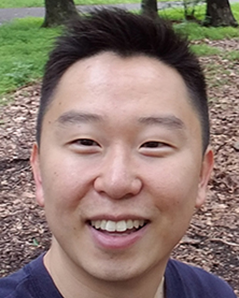
Jong Park, PhD
The animal models currently available to study gas-exchange functions in vertebrates are limited. Jong Park, PhD, a postdoctoral fellow in the Weinstein laboratory (Section on Vertebrate Organogenesis), presented the zebrafish gill as a new model for studying gas exchange in the vascular endothelium.
Dr. Park showed that the externally located zebrafish gills provide a unique model for studying gas exchange because they are optically clear and experimentally accessible, unlike mammalian lungs. Using single-cell RNA sequencing of dissected adult zebrafish gills, Dr. Park found that many specialized lung cell types are conserved in gills, and he identified a novel vascular endothelial cell subtype. In situ hybridization revealed that these novel cells are localized to the highly vascularized distal tips of the gill filaments where oxygen exchange takes place, suggesting an important role for these cells. As Dr. Park continues to characterize these novel endothelial cells using transgenic lines, he hopes to uncover their role in zebrafish models of lung diseases, such as acute respiratory distress syndrome, chronic obstructive pulmonary disease, or even COVID-19.
New Animal Models Could Aid Intellectual Disability Research
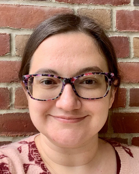
Rachel Cosby, PhD
Mutations in a Thanatos-Associated Protein (THAP)-coding gene called THAP7 could be responsible for some forms of intellectual disability (ID), according to DNA analysis conducted by Rachel Cosby, PhD, a postdoctoral fellow in the Macfarlan laboratory (Section on Mammalian Epigenome Reprogramming). Dr. Cosby utilized this insight from human patients to develop animal models of ID.
ID affects about two percent of individuals worldwide and is characterized by an IQ of less than 70, in addition to major skill impairments. The THAP7 gene is conserved across vertebrates, which enabled Dr. Cosby to observe mutant THAP7 phenotypes in a zebrafish model. Loss of THAP7 function led to reduced longevity in zebrafish, confirming the significance of THAP7 for organism health. Dr. Cosby also created a THAP7 knock-out mouse and will characterize the phenotypes of the mouse to evaluate its potential as a model for ID. Dr. Cosby plans to elucidate the unexplored functions of THAP7 to further uncover the pathology of ID induced by this mutation.
Demystifying Ribosomal DNA Repeats

Paul Atkins, PhD
Tandem repeats are an important, yet mysterious characteristic of ribosomal DNA (rDNA), the DNA that encodes the RNA strands critical for ribosomal function. Across species, some rDNA sequences are highly conserved while others are variable. Paul Atkins, PhD, a postdoctoral fellow in the Levin laboratory (Section on Eukaryotic Transposable Elements), is developing S. pombe (a type of fission yeast) as a model organism in which to study rDNA tandem repeats.
Dr. Atkins designed a pipeline to compare yeast genome datasets and created a novel method for assembling rDNA repeats from long DNA sequences. He compared 95 strains of S. pombe and S. kambucha and found a five-fold range of variation in rDNA repeat numbers. To explore the underlying mechanism of the variation, he performed a genome-wide association study and identified differences in genes that are involved in DNA maintenance, replication, and homologous recombination. Dr. Atkins plans to further investigate the role of these newly identified genes in regulating rDNA repeat number.
